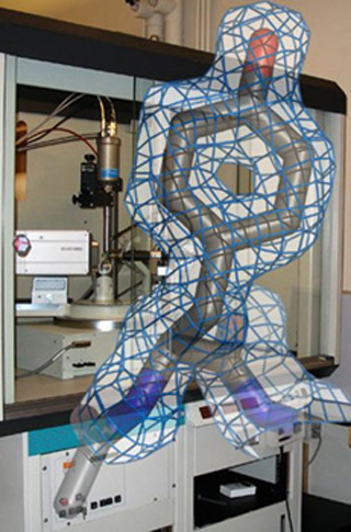
Molecular model of amino acid tyrosine overlaid on photograph of xray diffractometer
Description:
Molecular model of the amino acid tyrosine with experimental electron density in front of an X-ray diffractometer at MIT. The tyrosine is part of the crystal structure of phosphoglycerate mutase from M. tuberculosis. See Mueller, P., et al. Acta Cryst D61 (2005): 309-315. (Figure and photograph by Dr. Peter Mueller.)
file
183 kB
Molecular model of amino acid tyrosine overlaid on photograph of xray diffractometer
Alt text:
Molecular model of amino acid tyrosine overlaid on photograph of xray diffractometer.
Caption:
Molecular model of the amino acid tyrosine with experimental electron density in front of an X-ray diffractometer at MIT. The tyrosine is part of the crystal structure of phosphoglycerate mutase from M. tuberculosis. See Mueller, P., et al. Acta Cryst D61 (2005): 309-315. (Figure and photograph by Dr. Peter Mueller.)

Course Info
Learning Resource Types
notes
Lecture Notes


