Course Description
Did you know that we have approximately 2 meters of DNA packed in our cells, which are less than 10 μm diameter? Or that to replicate DNA it is copied at a rate of 70,000 basepairs per second by a cellular apparatus that coordinates at least six different enzymes? Or that microtubules form greater than 1 meter long …
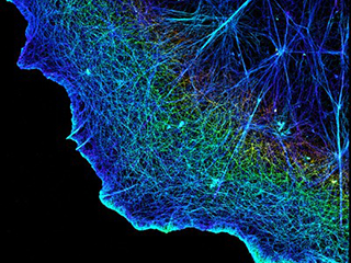
Did you know that we have approximately 2 meters of DNA packed in our cells, which are less than 10 μm diameter? Or that to replicate DNA it is copied at a rate of 70,000 basepairs per second by a cellular apparatus that coordinates at least six different enzymes? Or that microtubules form greater than 1 meter long “railways” upon which molecular machines transport cargo within nerve cells? In this course, we will explore how single-molecule imaging techniques capture the mega-cellular machines working in real-time.
This course is one of many Advanced Undergraduate Seminars offered by the Biology Department at MIT. These seminars are tailored for students with an interest in using primary research literature to discuss and learn about current biological research in a highly interactive setting. Many instructors of the Advanced Undergraduate Seminars are postdoctoral scientists with a strong interest in teaching.
Course Info
Instructor
Departments
Learning Resource Types