
Statistically-analyzed PET scan data superimposed on structural MRI scan
Description:
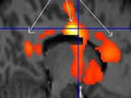
Statistically-analyzed PET scan data superimposed on structural MRI scan (front of brain is at right) shows areas in the anterior and posterior cingulate where panic disorder patients had nearly one third fewer serotonin 5-HT1A receptors compared to healthy control subjects. The lighter the color, the greater the difference between patients and controls. (Image courtesy of the National Institute of Mental Health.)
file
11 kB
Statistically-analyzed PET scan data superimposed on structural MRI scan
Alt text:
Statistically-analyzed PET scan data superimposed on structural MRI scan.
Caption:
Statistically-analyzed PET scan data superimposed on structural MRI scan (front of brain is at right) shows areas in the anterior and posterior cingulate where panic disorder patients had nearly one third fewer serotonin 5-HT1A receptors compared to healthy control subjects. The lighter the color, the greater the difference between patients and controls. (Image courtesy of the National Institute of Mental Health.)
Course Info
Instructor
Departments
As Taught In
Fall
2009
Level
Learning Resource Types
grading
Exams
notes
Lecture Notes
assignment
Written Assignments


