Wireframe Representation of a Fibroblast
Description:
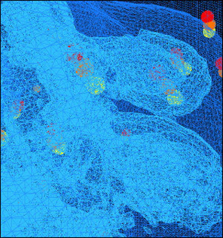
Actin-rich processes (light blue) in a transformed fibroblast protruding through extracellular matrix. The yellow spheres represent the centroids of beads added to the matrix to allow measurement of forces generated by the cell as it migrates through the 3D environment. The image shown is a wireframe representation of an isosurface rendering extracted from a time-resolved 3D series. (Image courtesy of Dr. James Evans of the Whitehead-MIT Bioimaging Center. Used with permission.)
file
233 kB
Wireframe Representation of a Fibroblast
Alt text:
Computerized image showing wireframe representation of a fibroblast.
Caption:
Actin-rich processes (light blue) in a transformed fibroblast protruding through extracellular matrix. The yellow spheres represent the centroids of beads added to the matrix to allow measurement of forces generated by the cell as it migrates through the 3D environment. The image shown is a wireframe representation of an isosurface rendering extracted from a time-resolved 3D series. (Image courtesy of Dr. James Evans of the Whitehead-MIT Bioimaging Center. Used with permission.)

Course Info
Instructors
Departments
As Taught In
Spring
2006


