
T1-weighted MRI Images (thumbnail)
Description:
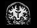
T1-weighted MRI images. Coronal section through the hippocampus. AD patients have shrunken hippocampi and enlarged ventricles relative to healthy age-matched controls. These changes result from cell dysfunction and cell death. (Image by Prof. Suzanne Corkin.)
file
6 kB
T1-weighted MRI Images (thumbnail)
Alt text:
Two MRI images side by side. One shows the brain of a healthy older adult, the other shows a brain of an adult with Alzheimer's disease.
Caption:
T1-weighted MRI images. Coronal section through the hippocampus. AD patients have shrunken hippocampi and enlarged ventricles relative to healthy age-matched controls. These changes result from cell dysfunction and cell death. (Image by Prof. Suzanne Corkin.)
Course Info
Instructors
Departments
As Taught In
Spring
2005
Level
Topics
Learning Resource Types
assignment_turned_in
Presentation Assignments with Examples


