EC.710 | Spring 2010 | Undergraduate

D-Lab: Medical Technologies for the Developing World

Projects

BabyTrackr

Labor contraction monitor for Nicaraguan hospitals

Team: Maysun M. Hasan, Grace Yao, Karina Isaak, and anonymous MIT students [LT] and [AB]

This content is presented courtesy of the students and used with permission.

1 2 3 Next »

Welcome to the Contraction Monitor team blog!

by Grace Yao

By the way, should we try to come up with a catchier name, maybe like Laborsaver or something like that? Just a random thing I thought of!

In any case, we are [AB], Maysun Hasan, Karina, [LT], and Grace Yao, and here is a little bit about the problem we are trying to solve and our goal for the solution.

In developing countries, such as Nicaragua, maternal health is very rudimentary in rural areas. Usually community health care workers make house visits to check up on the mothers, but they have limited knowledge and resources for proper maternal care. Also, mothers have to decide when to go to the health posts to address their concerns if any problems arise. When labor gets to a certain point, the ambulance has to be called to transport the mother to a hospital, which is often very far away. Often, it is hard for the mother to determine when she needs to call an ambulance. In worst-case scenarios, the mother doesn’t call in time for them to reach the hospital, and they must deliver the baby on the side of the road. To avoid such cases, we have proposed to develop a monitor to automatically alert different stages of labor based on contraction rate. The monitor should signal for when a contraction occurs, its length and rate, when the ambulance needs to be called, when critical attention is needed, when non-critical attention is needed, and when the baby needs to be delivered. The signals (in addition to the one for calling the ambulance) may aid health care workers in quickly identifying which mothers need attention at a birthing facility, helping them triage the patients.

Back to top

April 15 Post

by Karina Isaak

Hi team,

I just found this article which was published in May 2009.

Haws, Rachel A., Mohammad Yawar Yakoob, Tanya Soomro, et al. “Reducing Stillbirths: Screening and Monitoring During Pregnancy and Labour.” BMC Pregnancy and Childbirth 9, 2009.

It gives a review of the interventions used worldwide to prevent stillbirths.

Please, take a look at the section called

Monitoring in Labour  
Use of the partograph

starting on page 33.

They are talking about monitoring the contraction with a partograph, That’s kind of what we want to measure with our device. In the results section they say that even though they couldn’t find significant differences in maternal or perinatal outcomes with the use of partograph versus no partographs, it can still help in low-resource settings:

“Partographs may be comparatively more effective in low-resource settings, as the studies from Africa and Mexico in the Lavender review [140], as well as data from Southeast Asia [146] that showed reduced Caesarean section rates with use of the partograph and early intervention for slow progress of labour. The data from Southeast Asia and Indonesia also showed trends toward improved birth outcomes [146].” (Haws, et al., 2009, p.34)

Their statistical analysis was probably insignificant because of “unclear guidelines on the partograph use” (Haws, et al., 2009, p. 35). Therefore, building an intuitive device that is working with the partograph could possibly help to prevent stillbirths in developing countries.

What do you guys think?

Back to top

April 16 Post

Hey team,

Grace, I agree, we definitely need a more creative name… let’s brainstorm about it in class today. Here is a study comparing the EMG contraction monitor to a tocotransducer belt. EMG seems to be the better way to measure contractions.

Maul, H., W. L. Maner, G. Olson, et al. “Non-Invasive Transabdominal Uterine Electromyography Correlates with the Strength of Intrauterine Pressure and is Predictive of Labor and Delivery.” Journal of Maternal and Fetal-Neonatal Medicine 15, no. 5 (2004): 297-301.

Let’s first test the resistive flex sensor [LT] ordered and then start working on the EMG/EKG if the sensor turns out to be not reliable for measuring contractions.

Back to top

April 19 Post

by [AB]

Hey all,

Hope you’re enjoying the long weekend! I found another interesting article that shows how an external taco meter is just as good as an internal one, although the internal one uses pressure gauges. Just to confirm that we should make both an EMG and an external tocodynamometer. Anyways, I also think we could think of a better name. So I looked up LaborSaver, and apparently it’s already a product that allows workers to save money. How about laborhelper? Regardless, here’s the article.

Bakker, Jannet J. H., Corine J. M. Verhoeven, Petra F. Janssen, et al. “Outcomes after Internal Versus External Tocodynamometry for Monitoring Labor.” New England Journal of Medicine 362, no. 4 (2010): 306-13.

Here’s another article I found on how to make an embedded microcontroller and EMG:

Wu, Han-Chang, Chao-Hung Lin, Shuenn-Tsong Young, et al. “Monitoring Long-Term Uterine Contractions.” IEEE Instrumentation and Measurement Magazine 5, no. 2 (2002): 36-40.

I think it’s very similar to what Maysun sent out a couple days ago.

Let’s meet up some time (I think we’re agreeing for tomorrow 3 pm) to discuss specs and how to make both of these.

Back to top

Patents for Various Contraction Monitors

by Grace Yao

Apparatus for non-invasive monitoring of uterine contractions  
United States Patent 4989615

From Abstract: “An apparatus which includes a bladder element (10) which is at least partially filled with fluid, a belt-like element (12) which holds the bladder against the patient’s abdomen with some pressure, and a pressure monitoring device (18) which is connected to the bladder (10) to detect changes in the pressure of the fluid in the bladder (10) as the abdomen hardens due to uterine contractions.”

External uterine contraction monitoring device  
United States Patent 5070888

From Abstract: “An improved monitoring device for externally monitoring labor contractions preceding childbirth which does not require the use of a belt is disclosed consisting of a transducer removable assembly fixed to a base adhesively attached to the abdomen of the woman.”

Disposable tocodynamometer with self-adjusting bellows  
United States Patent 5224490

From Abstract: “A non-invasive, disposable, self-adjusting tocodynamometer (10) for monitoring uterine contractions of a patient during pregnancy, labor, and delivery The tocodynamometer includes a pressure-sensitive, fluid. filled bellows (20) responsive to changes in the hardness of the uterus during contractions The bellows has one face which projects into the patient’s soft tissue in the abdomen and adjacent the uterus, rending the tocodynamometer sensitive even for obese patients. A plate (22) supports the bellows and provides structure for attaching the tocodynamometer to the patient. A wall (30) formed on the plate receives the bellows as it is compressed during use. A conduit (14) connects the bellows to a pressure transducer (12) which, in turn, is connected to a monitor (16). The bellows, conduit, and pressure transducer form a closed system containing the working fluid.”

Back to top

Retroactive Post

by Grace Yao

This is where we were 5 days ago, before the meeting with Jose:

Below is a list of Ob/Gyn’s in Boston that I grabbed from thecityofboston.com. I think we’ve pretty much decided on using the simpler on/off indicator as opposed to measuring force with varying intensity. So what we really need to find out now on the medical side of things is:

What are the times we have to measure to know the things we want to indicate, which are

  1. when the mother should go to the hospital
  2. when the mother needs attention but not critical
  3. when the mother needs critical attention
  4. when the doctor needs to be there to deliver the baby.

Then the 5th thing we’re having a light for is an indicator of when she’s actually having a contraction, which is pretty straightforward.

Other things we have to figure out:

Materials: what kind of material for the squeezy part? The wire connecting the ball/wristband should have a covering too right? What is the wristband going to be made out of? We want to look into refractive materials so that we can just use one light to light up a lot of the wristband.

Smarts: Microcontroller? Something else? Maysun, can you maybe summarize what you talked about with Paul?

User Interface: How much information should the mother have? How are we going to display it?  
(Number/graph/both?) Do we need to design a radio component too? Would sound be better?

Acker, David B MD - Brigham Women’s Hospital

75 Francis St Ste ASB1

Boston, MA

(617) 732-5445

Etkin, Masha J MD - Vincent Obstetrics/Gynecology Associates

32 Fruit St Ste 4E

Boston, MA

(617) 726-1753

Gomez-Carrion, Yvonne MD - Beth Israel Deaconess Women’s

330 Brookline Ave Ste KS205

Boston, MA

(617) 667-2952

Johnson, Kim M MD - Kim M Johnson MD

500 Brookline Ave Ste E

Boston, MA

(617) 732-6399

Moody, David B MD - David B Moody MD

45 Francis St

Boston, MA

(617) 732-6389

Perkins, Rebecca B MD - Rebecca B Perkins MD

85 E Concord St

Boston, MA

(617) 638-8000

Rodriquez, Elisa MD - New England Medical Center

860 Washington St Ste 2

Boston, MA

(617) 636-6114

Taylor, Faye - Boston Medical Center

91 E Concord St Ste 6

Boston, MA

(617) 414-5461

Wakamatsu, May M MD - May M Wakamatsu MD

55 Fruit St Ste 148

Boston, MA

(617) 726-2000

Yum, Mimi MD - Mimi Yum MD

330 Brookline Ave

Boston, MA

(617) 667-0478

Back to top

Team Info, Revised Problem Statement, Some Early Brainstorming Thoughts (Pugh Chart, Design Specs)

by [LT]

Hi! This is also sort of a retroactive post. We are the Contraction Monitor team!

When a woman goes into labor, clinics are not equipped to monitor her contractions- an important indicator of her stage of labor and any complications. Health workers currently rely on their hands on the woman’s stomach to time the contractions, fetal heart rate, and feel for the relative intensity. This can be a time and labor intensive method, especially when 8-10 women are in labor at the same time in the same room. We have proposed to develop a monitor to automatically alert different stages of labor based on contraction rate. Additionally, it will give the relative intensity of the contraction and output a graph with the contraction frequency. The monitor should signal for when labor has started, when a contraction occurs, its length and rate, when critical attention is needed, when non-critical attention is needed, and when the baby needs to be delivered.

Some thoughts from early brainstorming:

Should we be wary of giving to much info to mother? consider external monitor

Distributed beforehand? when starts feeling contractions, puts it on, tells her when she needs to go to hospital based on timing (how much time she has left, etc.), then switches to more accurate mode with wireless broadcast. Have extra mode or signal for when to go to hospital?- Since we’ve revised our problem statement, this is no longer a priority need!

Talk to potential users about what signals to use, and whether she rather know or now know, and what would stress her out

The iPhone® app contraction monitor has start time, end time, duration, and frequency. Has a timer, and history. Another app gives the graph.

Thought for Ball contraction monitor: info sent to doctor’s phone.

See below for our Pugh chart and Prelim Design Specs!
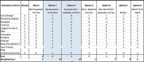
Pugh Chart

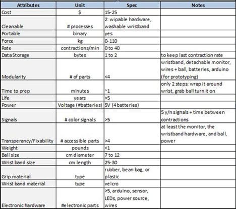
Design specifications

Back to top

Pitch

by [LT]

A first attempt at a pitch!

In developing countries, such as Nicaragua, clinics are not equipped to monitor a woman’s contractions while she is labor- an important indicator of her stage of labor. Health workers currently rely on their hands- which can be time and labor intensive, especially when 8-10 women are in labor at the same time in the same room. Our innovation will give health workers the ability to monitor contraction rate and alert them to the different phased of labor.

When the woman is picked up from her home, the health worker places a band with a monitor around her wrist. Attached to it is a ball that the woman squeezes when she has a contraction. One of five signals will light on the wristband to indicate what the woman is experiencing, such as a contraction or complication that needs immediate attention and a contraction rate will display on the monitor. Especially when only one health worker is present in the ambulance truck and must drive, the visual cues will help health professionals know how to respond immediately.

Back to top

Re-direction of our Project

After meeting with Jose for feedback on our project, we began to change directions (and revise our problem statement). Instead of working on a squeeze ball monitor alone, we’re also prototyping a belt (testing various sensors) and comparing that with the squeeze ball. Ultimately, it will become a behavioral study.

Summary of Our Meeting:

We went over what we were planning to do and Jose began to push us to try the belt idea. He’s concerned that since the woman control the input with the ball, she can either be too urgent and squeeze it too many times to go to the hospital or not squeeze it enough. He encouraged us to do a behavioral study comparing the ball and belt to see the correlation or accuracy of signals (if we find the ball is just as good and even cheaper, then we go with the ball, for example). Basically he told us to explore both options. We also discussed technical details and the sensors. He thought what we were doing was…settling for too little and we could do more. He encouraged us to explore more bio sensors, homemade EMGs (suggested by Maysun), and other flexi sensors and to really talk to Professor Frey. Maybe even look at making our own sensors if we think we can make them cheaply.

We went over what the problem really was and it’s actually that they don’t use any monitors and need something in the hospital (not necessarily at the home to tell them when to go). The current are too expensive, so we need to make it affordable. He told us to contact Ken Endo at the biomechatronics lab, and sent us an email introducing us to some Nicaraguan contacts I think.

So:

  1. Explore homemade sensors and other sensors, continue contacting people, figure out the smarts with a computer first
  2. Design: make prototypes (ball and belt) and turn comp into smarts with micro controller
  3. Clinical: test it out, behavioral studies

Back to top

1 2 3 Next »

<< 1 2 3 Next »

The Smarts, and How We Got There.

by Maysun M. Hasan

Hi guys,

This is my first post so I guess I’m going to back track a little. Please excuse any spelling mistakes… I am the worst speller in the world. The first thing I did when I found out about this project is call my mom… I know, kind of lame but whatever. She is an OB/Gyn and she has worked in many different types of settings, the most relevant for us being in the labor room at a rural Bangladesh clinic.

She told me that during labor, healthcare professionals monitor a woman’s contraction rate, and cervical dilation to judge the progress of labor. A woman can be in labor for many hours but only when her contractions are close together and intense, and she is at least 10 cm dilated, is she close to delivering. My mom had said that once the contractions are close together (approximately 3-4 minutes apart), a doctor should come to examine the patient, and see if everything is alright. The doctor needs to see how much the woman is dilated. If the woman is not dilated enough, she should be given drugs to lessen the contractions for a few more hours. The doctor should also check the position of the baby, and reposition it if it is necessary. All this should be done at approximately this stage of labor, because if the doctor checks later, it might be too late for an easy intervention. So one thing our device should do is indicate when contractions are a 3-4 minutes apart (we should find a firmer number in literature) so that the doctor knows they need to be around.

My mom had also mentioned that fetal monitoring during a contraction is also very important, especially to detect early fetal distress due to fetal hypoxia, and metabolic acidosis. What normally happens is what is called mirroring. As the uterus contracts, the fetus goes into distress and its heart rate drops, but should come up immediately after the contraction has ended. If it doesn’t, and the heart rate stays down after the contraction has ended (called late deceleration or uteroplacental insufficiency) it indicates a fetus’ inability to respond to stress, most likely due to lack of oxygen and invention by the doctor is necessary. Thus knowing when a women is having a contraction, from start to end, is important information our device should delivery. My mom said, the current method of doing this, in ill-equipped settings, is with a stethoscope. When a woman is having a contraction, the muscles harden, making it harder to hear the fetal heart beat. Thus the heart rate starting from when you can hear it indicates late deceleration. This hearing method is not optimal, so our device should address this.

Lastly, my mom told me that abnormal change in contraction rate is also important data. For example, if the contractions are going from 10 to 9 to 8 minutes apart to all of a sudden 10 again, something probably happened. This is most of the time not critical, like the mother needs to go to the bathroom or something, but either way she needs attention. So our device should show when the contraction rate indicate that non-critical attention is needed.

And lastly, my mom told me about uterine inertia. This is when the uterus atrophies, and if this happens, the mother should be rushed to a C-section. An indication from contraction rates for this is if the contractions go from 4 to 3 to more than 30 minutes apart. Thus signaling when the uterus atrophies is something our device should do.

A lot more long winded than I wanted to write but whatever. This is just what we believe our device should do along with indicating the actual rate of contractions, and possibly intensity and duration.

We plan on doing this with the Arduino. We talked to Paul about this. We want our device to be relatively cheap so all the women in the labor room can have one, so I was originally not loving the >$20 Arduino. A microprocessor is not really necessary here since only solid logic is involved. I was thinking we could make a new chip that would do only this. It would be cheaper than and not as bulky as the whole Arduino. However, Paul got me to change my mind. The microprocessor itself is very small and cheap (~$3), so we could program in one place and attach to the device somewhere else. This is a lot better than making a new chip (which is easy to design but probably a pain to manufacture).

So we know approximately what we want, and we know how to do it, the next step is doing it. We also need to know the input to our device: squeeze ball, EMG, flexi-force… etc, but that’s a different story. Sorry about the novel guys.

Back to top

Updates 4/24

by [LT]

Updates

  • Karina and [LT] made two switch prototypes (massacred a bear and used his leg for a big red button, attempted to stick a switch in the tiger- his guts were everywhere, but he’s not bandaged)
  • [LT] and Karina met with Paul Saturday (today 4/24) to begin logic for switches and work on logic for data storage (what information we want to collection, display and how)
  • Our resistive flexi strain gauges have arrived!
  • Paul can order us a display for our prototypes
  • Maysun working on EMG sensor p.m. Sunday (tomorrow 4/25)
  • [AB] and Grace, please feel free to reply to the team with updates on information on posts on blog!
  • Maysun working on EMG

Meeting Agenda (4/25)

  • Update on sensor prototypes – Maysun, Karina, [LT] ~ 10 min
  • Update on research – [AB], Grace ~ 10 min
  • Decide deadlines for research, code and prototypes ~ 15 min
  • Decide on display ~ 20 min
    • Graph or dips with LED
    • Just numbers and signal lights
    • Computer

Gantt Chart review ~ 5 min

List of to-dos and to-blogs ~ 5 min

Total ~ 1hour 5 min

Back to top

Research

by [AB]

Hi all,

Here were the three topics I just researched for discussion at the next meeting:

  1. Complications during pregnancy–uterus twitching
  2. What else occurs when contraction happens, how flexiforce can be used
  3. How to make a partogram

For (1) I found a helpful Web site that lists all the disorders that occur during pregnancy. All high-risk patients should already be monitored closely, and those include: women with chronic hypertension, diabetes, lupus, incompetent cervix, placenta previa, preeclampsia, premature birth, recurrent miscarriages, and thyroid disorders. I think the disorders we should worry about are connected to the uterus in some way and might give false positives when using the belt we’re building:

A) Fibroid Tumors: They are usually undetected non-cancerous masses that occur in the uterus, that go easily undetected in the third trimester of labor. There are four different types of fibroids in the uterus, subserosal, Intramural, submucosal, and Pedunculated. The one that might affect the size of the uterus and is the most common is the Intramural Fibroids, because they develop within the uterine wall and makes the uterus feel larger than normal. This causes pressure and pelvic pain. Another one that could also grow outside the uterus is the Pedunculated fibroids. This enlargement might be confused with the pressure that a contraction makes if we use the flexiforce so we should see if we can use another type of monitor, such as the partogram, which measures the cervix dialation, can be used alongside the flexiforce belt.

B) Placental Abruption: The placenta carries all the nutrients and O2 to the baby in the uterus from the mother. There are two extremes to this disease. Minor abruption may irritate the uterus and cause induced labor, but usually the baby is safe. The other type is Massive abruption, which may result in early labor and delivery, fetal intolerance of labor so C-section is required. There should be a way to measure this abruption. The causes of this abruption are usually unknown but could be caused by injuries to abdomen, use of drugs, high blood pressure, or chronic diseases such as diabetes. All pregnant women with these complications should be closely monitored already, but if it happens while the woman is in the hospital, I think we should have a way to detect it, I believe that slowing down of contraction rate might not be the only way to tell if the placenta has ruptured. After some more research, I found that there is usually pain in between contractions, so we should have some sort of button or monitor of intensity of pain between contractions to check for complications as well? I don’t know how to do this without having an honor code for the women because they are the ones in pain. There is also vaginal bleeding so that might be easier to detect and then there is abdominal pain that remains constant throughout labor, which should not be confused with increase rate of contraction.

C) Uterine twitches: A normal contraction will occur by shortening and thickening of uterine wall muscle fiber. The contraction starts at the top of the uterine wall and waves down to the bottom. When “true” labor begins, women feel “false” labor pains, or a Braxton Hicks contraction, which is irregular uterine contractions that occur in the second or third trimester of pregnancy. They’re the body’s way of getting ready for the “real thing.” They feel like a tightening in the abdomen that comes and goes. They do not get closer together, do not increase with walking, don’t increase how long they last and don’t feel stronger over time. Here is a table that doctors ask their patients over the phone apparently:

CONTRACTION CHARACTERSTICS FALSE LABOR TRUE LABOR
How often do the contractions occur? Contractions come at regular intervals and last about 30-70 seconds. As time goes on, they get closer together. Contractions come at regular intervals and last about 30-70 seconds. As time goes on, they get closer together.
How strong are they? Contractions are usually weak and do not get much stronger. Or they may be strong at first and then get weaker. Contractions steadily increase in strength.
Do they change with movement? Contractions may stop when you walk or rest, or may even stop if you change positions. Contractions continue despite movement or changing positions.
Where do you feel the pain? Contractions are usually only felt in the front of the abdomen or pelvic region. Contractions usually start in the lower back and move to the front of the abdomen.

When twitching occurs, there is no wave-like movement of the contraction, so there is a distinct difference between the two. The twitches are more like a spasm, and also they occur in women who are not pregnant as well, and do not cause pain so there is another difference between contractions and twitches/spasms. One last little tidbit, which you guys might know already: women in the US go to the hospital with contractions occur every five minutes for an hour.

  1. I am still researching what else occurs during contraction other than water breaking, not in all cases, cervix dilation and random bowel movements that women can’t control.

  2. I think we should try and make partograms, they’re pretty easy and can monitor dilation, fetal heart rate, duration of labor and vital signs. That way delay or deviations of labor can be detected more easily than depending on contraction rate deviations. It also shows a numerical record of features such as urine output and the volume and type of intravenous infusions (including oxytocin drips):

Image removed due to copyright restrictions. Please see image and explanation of a partogram at http://www.2womenshealth.com/Childbirth/Partogram.htm

There are two-hour partograms and four-hour partograms, but I think the best one is to do a two-hour study (approved by WHO, because it increases the need for intervention without improving maternal or neonatal outcomes compared to four-hour partogram) and when the labor looks slow, the doctor will do something about it. Doctors say that a rate below the average (1 cm/h) shows a slow labor. First, they rupture the amniotic sac and then give oxytocin to stimulate contractions and labor. Partograms must be used along with the belt because when doctors stimulate contractions, careful monitoring should be made to ensure that contractions do not exceed one every two minutes. If this stimulation doesn’t help, then the doctor will perform a C-section, only after the mother’s consent.

I think the nurse or health official needs to keep track of this though, which may be a problem if there are more than five women in the ER. There is also clinical pelvimetry, which is used to monitor the size of the birth canal by means of systematic vaginal palpation of specific bony landmarks in the pelvis and the estimation of the distances between them. Internal pelvic diameters are not accessible, so usually midwives or a doctor will infer. This occurs at the first prenatal check-up (do they have those in Nicaragua??) and they’re recorded as adequate, borderline, or inadequate, rather than in cm or inches.

Maybe that might be a better idea than partogram? However, I think that we should still make a chart for the partogram and see if it’s intuitive enough for the doctors to use…

Anyways that’s what I’ve researched so far…will keep you guys updated on anything else i find. Sorry for the long post!

Back to top

Quick Question

So I know I couldn’t go to the meeting with Jose which may have been where I missed this, but whatever happened to the part about helping mothers know when to call the ambulance? I thought that based on what Ryan said about his personal experience and on one of the problems we had identified in Nicaragua, which was not having babies on the side of the road, we were going to make a home-use component too. Are we not doing that anymore? It seems like this is purely for use by health workers at the hospital - which is okay I guess; I just would like someone to justify why.

Back to top

Button Logic Pseudocode and Visuals

by Grace Yao

So this is what we talked about in class today, besides looking at the button models [LT] and Karina made and eating M&M’s from Anna!

Here’s a vague idea of how we want the button to work…

boolean down #true = button is pushed down

# 3 states: notContracting, checking, contracting

if state == “notContracting” and down:

   state = “checking” # change the state

if state == “contracting” and !down:

   state = “checking” # change the state

if state == “checking”:

if (down for >= 5 seconds): # 5s so nothing will be counted mistakenly. need to somehow start timing when down changes from t->f or f->t?

   state = “contracting”

elif (!down for >= 5 seconds):

   state = “notContracting”

Visuals should include these:

  1. The display should have 2 rows of numbers, top row = duration of contraction in seconds, bottom row = duration of time in between contraction in minutes; these numbers would preferably be offset from each other in a zigzag, but we’ll see depending on what the monitor looks like.

  2. The two little number counters should show 

    a) Rate (how many contractions in the last hour) and 

    b) Time since the last contraction

  3. Three differently-colored LEDs. 

    a) Contraction button is pressed, 

    b) when the baby is about to come, aka when rate = 10 contractions/hr and time in between contractions = 5 minutes, and 

    c) When there may be a complication -> steady if not urgent, blinking if urgent.

  4. Maybe a row of LEDs to show intensity?

Meeting 4/28- What we’re Up To!

by [LT]


Gantt Chart

Hi Everyone,

The Gantt indicated when each step should be finished!

A new quick capacitor idea: Maysun will work on putting one with conductive metal, a belt to constrain it, wire, and rubber from bicycle inner-tube from D-Lab to prove that it might be more viable than the EMG and pick up on contractions by change in capacitance. She suspects that EMGs might not be robust enough and the electrodes must be disposable.

The capacitor will work off displacement of the muscle and be slightly pressed on the woman’s stomach. When the muscle tenses, the plates will close up, and we should observe some change.

Grace and I will be finishing up the general code and the screen to display contraction data has been ordered by Paul and should be coming soon. Karina and I will finish the actual button on Sunday. Grace and [AB] will be starting up the poster. Please comment to this if I forgot something!

Back to top

EMG Plan

by Maysun M. Hasan

Hi guys,

So I should have posted this about a week ago, but my computer died and stuff happened so it never got posted. This is the plan for the EMG.

The Electromyography (EMG) is a technique of recording the electrical activity in muscles. So the principle theory is that uterine contractions are caused by a series of action potentials that will cause a voltage change that can be detected, similar to how an ECG works. So how should we go about making an EMG? First we need a way to detect electrical activity. We can do this with a differential amplifier. The voltage difference is measured from two electrodes, which are placed relatively close together on the surface of the mother belly, (on the uterus). They are compared to ground, which is place on the mother’s hip, where there is no muscle activity. The Diff Amp should output a signal like below, which is an EMG for a bicep:

Image by MIT OpenCourseWare.

This signal has been high and low passed filtered, which is something our circuit needs to do. The signal should then be rectified so that only the positive part of the signal is kept, like below.

Image by MIT OpenCourseWare.

Finally, we need to smooth our signal out so that we form an “envelope”, as seen below.

Image by MIT OpenCourseWare.

So our overall EMG system is described in the flow chart below.

This is our plan. Let’s see how it goes. I think getting the Diff Amp to give a signal, which is really small, and filtering all the noise is going to be the hardest parts. Since we lack a pregnant women, we will be testing our system on our arms. [AB] and I tried to get electrodes from MIT Medical, but ended up getting the run around and failed miserably in our quest. But fortunately my friend Ankit said that he could get us electrodes. So all is well, I hope, at least we’ll see when we start building.

Source: (PDF)

Back to top

Playing Around With Different Materials, Toys and Arduinos

by Karina Isaak

Here are some pictures of our experiments with different sensors (buttons) and materials for the contraction monitor ball, that the women in labor squeezes if she is having a contraction.

Our favorite is the big red button (picture 1) put into a latex glove which is filled with cotton (pillow stuffing). It is soft, comfortable, cheap, easy to produce, robust and reliable.

We also started programming the Arduino to record when the ball is squeezed, indicating that the women are having contraction. Grace posted the pseudo code that we are working on.

The next step will be to connect it to the display and get everything running!


<< 1 2 3 Next »

<< Previous 1 2 3

Cloth Patch for Monitoring Contractions!
by Karina Isaak

Hey guys,

I found a pressure sensor matrix that we could adapt to a pressure sensor patch on the stomach of the women in labor on this webpage (see below). It gives instructions how to build a pressure sensor by using two pieces of cloth and conductive thread! If I understand it right, you just have to sew everything together in the explained way and connect it with external pull-up resistors! They even provide the Arduino code. Click through the pictures on the top, all steps are explained in detail and a video is provided in which you can see how it works.

http://www.instructables.com/id/Pressure-Sensor-Matrix/

We should try this! Maybe we can make it as a sticky patch.

What do you think?

Back to top

Home Stretch
by Grace Yao

It’s crunch time now! MIT Museum 1-minute pitches this Saturday, and 15-minute presentations in class next week.

We’ve finally chosen a team name: Team Partayuda, from the Spanish words for “birth” and “help.” So here’s what’s up, as far as I know…

Maysun is working on the breadboard and programming for the pressure sensor - a capacitor, with rubber in the middle, and [AB] is going to make it. We tried making the EMG last week, but it did not work out.

Karina found the cool conductive thread project that she just posted, so she and [LT] are going to try to make that happen; [LT] is also working on Arduino code for the button.

So in total we should have 3 different ways of input to test (which may or may not happen before class presentations): the capacitor, the conductive thread, and the button…I think right now the button is the closest to being finished.

I’m going to work on the processing code to take the input and provide an output of the results we want to show. However, we don’t have the monitor yet, so this may end up just being displayed on a computer. [AB] and I are going to put together the poster, including the problem statement, our solution, Pugh chart, Design Compass, and design specs among other things, while [LT] is going to make a slide for the museum pitch.

Busy week, but we’re almost there!

Back to top

Response From Nicaragua
by [LT]

Hi All,

Our contacts in Nicaragua have kindly responded to our questions with very interesting information that has really defined our problem. See their responses below.

  1. The women know about when to arrive at the hospital. They receive information when they receive their prenatal checkup. Births can happen during transport due to the distances involved. Women don’t have ready access to an OB/Gyn and subsequently need to travel up to 4 hours to receive specialized attention.
  2. The monitoring is crucial in knowing the condition of the mother and the fetus.
  3. We use a medical method in which we place a hand over the patient’s abdomen for 10 minutes and perceive the contractions. We have immense difficulties because of the large number of patients. There are only 2 doctors per shift for 15-25 patients which need to be monitored every 4 hours and apart from that, we need to attend to births and do cesareans and fill out a stressful amount of paperwork. On occasion, we don’t manage to monitor all the patients and are limited to just asking if they have had pains and with what frequency. To listen to the fetal heart rate, we use a stethoscope. Often, we don’t listen to it because we have to move them around to listen to it with a fixed ultrasound which takes up more time.
  4. To give you an idea of how difficult it is to monitor all the patients, before every shift, we pray to God that [the room] is not too full.
  5. To see if it would cause the mother stress, it would be necessary to conduct a study, and that depends on various factors, but for us it would be very valuable information.
  6. Don’t know if it will be effective or not. For that, we’d have to try it, but I think that for the level of education, it would be better that they didn’t participate (I think he means that due to their lack of education, they wouldn’t do it right- they shouldn’t have control of the input of the monitor).
  7. Contraction control is crucial. We do it every four hours when the expectant mother doesn’t have any labor problems and each hour when she is in labor. This last case we control the labor through the partogram.

Back to top

Instructable
by Grace Yao

I think the Processing code this girl wrote could be helpful to us right now (although our pulse ox code might already do the trick for now); the entire Instructable might be useful in the long run for future work.

http://www.instructables.com/id/Fabric-bend-sensor/

“Using conductive thread, Velostat and neoprene, sew your own fabric bend sensor.

This bend sensor actually reacts (decreases in resistance) to pressure, not specifically to bend. But because it is sandwiched between two layers of neoprene (rather sturdy fabric), pressure is exerted while bending, thus allowing one to measure bend (angle) via pressure. Make sense? Watch below:”

By the way, our team name is now Babytrakr!

Back to top

Pennies for Your Muscles
by Maysun M. Hasan

Hey,

So I totally suck at blogging!!! I have a lot to say about the progress of our device that has happened in the last week. The last time I wrote, I had a plan for the EMG and I was about to build. Well I built it and realized that this was not the best way to do it… This is the schematics for it anyways.

Image by MIT OpenCourseWare. Adapted from circuit designed by Ankit Gordhandas, Irena Hwang, and Vinay Ramasesh.

This is the differential amplifier stage I described earlier. I built it, along with super cheap, super cool electrodes :) (See below)

Pennies as electrodes

Yes… they are pennies that I soldered to wires… but they work so whatever. So I build the circuit with LM741’s (the most typical op amp you can get)…and… it didn’t work. I know it was able to take the difference between two signal… but not muscle signals. So I did some research and found out that because it is such a tiny signal… and better op amp is necessary. So I used LT1637’s for the buffer and a LM358 as my amplifier. And… it didn’t work. After debugging though… I found my signal… But!!! It was tiny and very noisy!!! Also it was not the same shape as we thought it would be. It marked when the muscle started to contract, but not the whole contraction. This signal was so inconsistent that I felt it would not be the best for our prototype. That is why EMG was out… but what now… we have no input… Well this is where the capacitor idea that is mentioned in a few of the blog entries comes in. Sorry for the weird timeline… I am horrible at posting.

Back to top

Convo with Dr. Acker
by [AB]

Thanks Mays for reminding me to post also! Sorry this is very late and happened 2 weeks ago but here’s how the conversation with Dr. David Acker from Brigham and Women’s Hospital went:

  1. What do you use to measure contraction?
  • he measures the frequency, duration, and intensity of each contraction

There are 3 subjective ways to measure:

  1. ask woman how much it hurts and to record frequency of contractions on paper
  2. place monitor on abdomen and when contraction causes stomach to rise the pressure sensitive device will monitor by graphing on a slowly moving strip of paper (can’t record the intensity of the contraction with this device, however.)
  3. membrane ruptured and tubing placed through vagina into uterine cavity—intra-amniotic pressure measured and this is the most precise way to measure contractions but it’s invasive and uncommonly used
  1. How does the tocodynamometer work?
  • Measures contraction rise by pressure sensitive sensor (device is like a piston)
  1. When do you know baby is coming?
  • Contractions occur every 5 minutes lasting 1 minute each for the past hour, then the baby is going to come soon
  • There is monitoring of the woman even a week before the baby comes
  • Labor is not defined by the contraction rate, it is defined by the contractions leading to a cervical change (that’s where the partogram comes in)
  • Rock hard cervix until contractions loosen it for opening
  • Fetal heart rate response indicates complications (our device will notify the doctor when the contraction ends so he can check the fetal heart rate, it will not have a fetal heart rate monitor…yet?)
  • On average, the fetal heart rate shouldn’t change, but when fetal heart rate increases → reassuring because indicates an acid/base balance
  • When fetal heart rate decreases → various patterns so doctor needs to determine which pattern happens to see what is wrong
  1. What smarts are the best out of EMG, flexiforce or push button?
  • In the US, Dr. Acker doesn’t believe a push button will be effective because we’re simply replacing a pen and pencil for the women to a ball. However, he thinks it might help if they are in lots of pain and don’t wish to record their contractions.
  • He thinks the rubber pressure sensor is a very cool idea, however, the abdominal muscle or rather skin on top of the abdomen doesn’t harden so the pressure sensor should be something that notes rises in abdomen (~1 cm rise, Maysun’s mom also said the same thing)
  • Women’s abdomens are different so an external sensor needs to accommodate these variations
  • Thickness of abdominal wall also varies

We took all these answers into consideration when brainstorming for our prototype two weeks ago. As you can see in the post below, our prototype changed numerous times because we wanted to accommodate all the ideas and comments from doctors in Nicaragua and the US. Ultimately, we went for a pressure sensor that is different from a toco, but still measures abdominal rise for contraction rate to correlate to a hand squeeze ball. Dr. Acker also agreed to let us see his operation room if we needed some more ideas, so if the project continues we have access to his resources. He was very nice about answering questions and very interested in helping out in the future.

Back to top

Radio Awesomeness
by Maysun M. Hasan

Ok, it’s me again,

So, last time I told about the EMG circuit, which we rejected from our prototype because the signal was not what we expected and it was way too small and noise. I was reading up on other ways to do determine contractions (this was about 2-3 weeks ago), and discovered that EMG is not common method to detect contractions, even in the use. This is because there is also of other muscle interference, like fetal heart beat, maternal heartbeat, and maternal body movement artifacts. There is some cool research into additive filtering, and also how EMG’s are better in detecting when the baby is going to be delivered, but this research is not even applied here in the US, and what doctors need now is something that works. That is why we switched focus to a tocodynamometer, toco for short. This is basically a pressure sensor on the stomach that detects the upward movement of the uterus during contraction:


Courtesy Hesperian Foundation. Used with permission. From Werner, David, Carol Thuman, and Jane Maxwell. Where There Is No Doctor: A Village Health Care Handbook. Berkeley, CA: Hesperian Foundation, 2010, pp. 258. ISBN: 9780942364156.

We have been told that during a contraction, the uterus stiffens and moves about 1cm up. This is where the Toco comes in. It measures the pressure exerted by this upward force. After a long brainstorming section, we decided that a cool why to make a cheap and use to manufacture sensor was to make a capacitor. For a parallel plate capacitor, the equation is:

C= ϵ0 ϵr A/D

so as you squeeze the plates together, the capacitance changes, and all you need for the sensor are 2 conductive plates, which is locally available!!! I thought it was brilliant. To measure the difference in capacitance, I was going to make a simple LC oscillator, and have the sensor is parallel with the oscillator C. Thus, as the plates are squeezed together, the frequency of the oscillator changes, and we can plot the frequency changes over time. Here is the oscillator, the antenna in the picture is our sensor:

Image by MIT OpenCourseWare. Adapted from Ron Roscoe’s 6.102 Introductory RF Design Laboratory, Laboratory No. 4, Spring 2008.

I figured that there was about 10 pF variable capacitance, so in order to get a good frequency range, I was going to have two oscillators and mixer them and filter out all the frequencies I didn’t want:

Circuit design to filter unwanted frequencies

I know, a little technical… but it needs to be documented.

The mixer is below:

Image by MIT OpenCourseWare

Now!!! The really cool part of this is that these are all parts of a radio!!! A radio is cheap and there are already tons of mechanics who know how to work/fix them. The infrastructure exists and it would be easy to implement anywhere. I was so amazed with myself, I was telling a friend while debugging my circuit, and that’s when Velostat came into the picture. My friend saw what I wanted to do, told a much simpler and cheaper way to do this. So this eliminates our second prototype. Next time, Velostat and how it works!!!

Back to top

Logo
by Grace Yao

_
_

Team name in baby blue block letters next to baby’s footprints.

Karina and Nathan put together this logo last week for our poster - isn’t it cute?! Sort of makes the whole project feel more legitimate to me (not that it didn’t before, but having some sort of branding helps).

Back to top

Alien Mind Control!!!
by Maysun M. Hasan

Hi guys,

I feel I need to write this blog entry before the end of the term. So we decided at the end to use Velostat, a material I found through a friend, as our pressure sensor. So the technical description that 3M, the manufacturer of the material, provides is an “opaque, volume-conductive, carbon-impregnated polyolefin. Easily grounded, the electrical characteristics are not affected by age or humidity, and are suited for handling, shipping and storage.” I found this material at a site called lessemf.com, a site that, hilariously enough, specializes in materials to prevent alien abduction. Velostat is used to make into helmets to prevent alien mind control. But the real use is basically, it is the material used sometimes as anti-static bags for electrics. Electronics are packed in this stuff and often trashed afterwards.

It costs about $2 for a 1’x3’ piece. Now, what is Velostat? Basically, it is a material that changes resistance as you deform it, and it can be deformed by pressure, or bend. The resistance of the material untouched is about 28 K ohm, but as you bend or apply pressure to it drops. I hooked about a sq. inch of this up to a simple resister to make a voltage divider, high pass filtered it with a capacitor and resister, and voila, a very cheap pressure sensor.

Now working with it, I did find some problems. There is actually no stable baseline, but that’s where the high-pass filter helps. The problem is there is a definite limit to the filter, since our actual signal is very low frequency. But, this is not a big concern after filtering since we can threshold the sensor in the Arduino. Our second problem was that Velostat seems to be sensitive to heat and to sweat. Now this is a huge problem in our application. Fortunately, a piece of scotch tape around the material seemed to help. Lastly, since it is a flexible thin material, there is no easy way to attach wires to the material so there is often lose material. A cool thing, that a lady during our presentation at the MIT museum mentioned was that we could use conductive tape, like copper tape, but this adds cost.

To display the waveform the pressure sensor picked up, I used this code to make a simple oscilloscope with an Arduino (http://accrochages.drone.ws/en/node/90). With this display, I tested out different methods of arranging the Velostat to make it work better. First, I tried bending the material like this:

Arrangement of Velostat during testing.

Then I tried double layering it so to increase reaction. Both methods worked but doubling the layers was more sensitive.

So, after many iterations of the designing the input… ball, to EMG, to Capacitor, we finally reach Velostat. More stuff needs to be looked into, but it is definitely cool.

I would just like to give special thanks to [anonymous MIT student EZ]. I basically forced him to give me some Velostat to experiment with and graciously helped me to make the sensor work optimally.

Back to top

Presentation at MIT Museum
by Grace Yao

A couple of action shots from the museum presentations last week!

Back to top

More Action Shots PLUS Interested Physician from Brazil!
by [AB]

You probably can’t read the business card very well because the shot was taken from my iPhone® but Directora Terza Cristina Melo de Brito Carvalho is very interested in our prototype. She works at the Universidade de Sao Paulo and I’m going to e-mail her a copy of our poster. We should keep in contact with her and others interested for the future when we start making more prototypes. The last two pictures are from the EMG testing, and the few minutes that it worked before we realized it wasn’t grounded properly on grace’s arm. The rest are from the poster session (one with Amit’s daughter testing it out), and a picture of the actual poster. I can also upload a file version of the poster soon. Enjoy the pics!

Course Info

Instructor
Departments
As Taught In
Spring 2010
Learning Resource Types
Design Assignments with Examples